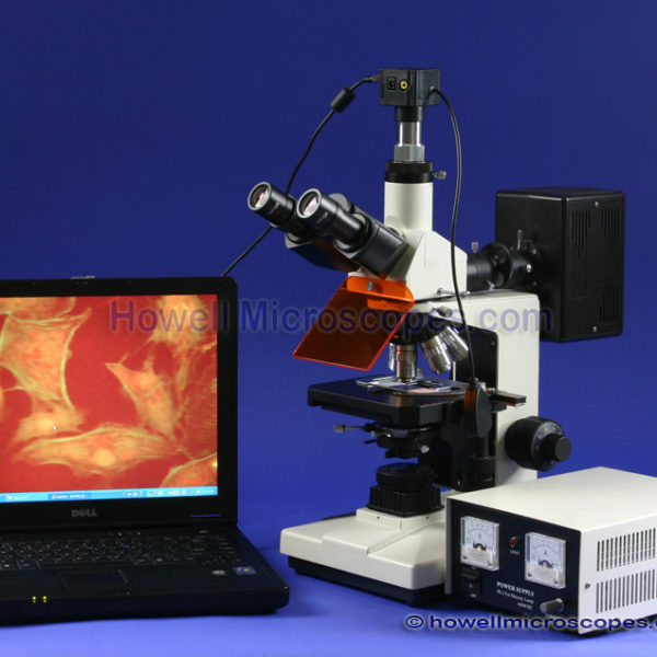
| Trinocular Photography Port |
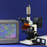
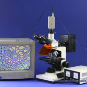
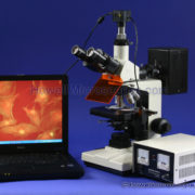
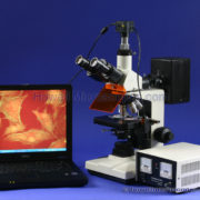
- Photograph Your Own Specimens!
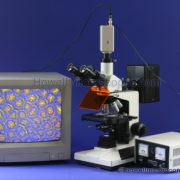
- Have Your Own Complete Digital Video Microscopy System!
- Capture Images With Your Computer!
- Videotape What You See Under the Microscope!
- Versatility – Included Trinocular Port Greatly Expands the Usefulness adding Photographic/Video Capability!
|
|
| Eyepieces and Magnification |
- Four Fluorescence Magnifications: 40x, 100x, 250x, and 400x.
- Two Fluorescence DIN Objectives: 25x (0.65 NA, 0.62mm Working Distance), 40x (Glycerin Immersion or can use Dry, 1.0 NA, 0.25mm W.D.).
- Optional (Not Included): 100x Fluorescence DIN Objective, 1.25 NA, 0.18mm W.D., Glycerin.
- The BrightField 4x and 10x Objectives can be used to Observe Fluorescence Images. For Higher Magnifications, the Special Fluorescence Objectives are used.
- Four BrightField Magnifications: 40x, 100x, 400x, and 1000x.
- Four Plan DIN BrightField Objectives: 4x (0.1 NA, 17.9mm W.D.), 10x (0.25 NA, 8.8mm W.D.), 40x (0.65 NA, 0.56mm W.D.), 100x (1.25 NA, 0.33mm W.D., Oil Immersion).
- Eyepiece Set Included: Wide Field 10x.
- Spring Loaded 25x, 40x, and 100x Objectives to Protect Slides.
- Reverse Facing Quintuple Nosepiece! A Better Design that allows Easier Access to the Slides since Objectives are Pointed Out of the Way!
|
|
| Head – Interpupillary Distance – Diopter |
- 30 Degree Inclined Trinocular Head (Easier to View than 45 Deg.).
- Adjusts to the Distance Between your Eyes: 53 to 75mm InterPupillary Distance.
- Diopter Adjustment to Correct for Your Specific Vision Needs.
|
|
| Condenser – Iris – Filters |
- Fluorescence Light Condenser with Condenser Movement Knob.
- Fluorescence Light Field Diaphragm with Centering Adjustment Knobs.
- BrightField: 1.25 N.A. Abbe Substage Condenser, Condenser Movement Knob, Iris Diaphragm, Swing-Out Filter Holder, and Condenser Alignment Screws.
- BrightField: Four BrightField Filters Included: Blue, Green, Yellow, Frosted White.
|
|
| Illumination |
- Fluorescence Light Source: Super High Pressure Mercury Lamp (100W DC). Mounted to the Rear and Enclosed in a Sturdy Metal Housing.
- External Power Supply for Mercury Lamp: 100W DC with Current Adjustment Knob and Volt / Amp Analog Gauges.
- The Arc of the Mercury Lamp is Easily Centered by using Two Lamp Positioning Knobs on the Lamp Housing.
- Epi-Fluorescence Illumination System: Blue and Green Excitation Fluorescence Filter Cubes.
- BLUE Fluorescence Excitation Dichroic Filter Cube: Excitation Spectrum 420 – 485nm, Emission Filter 515nm.
- GREEN Fluorescence Excitation Dichroic Filter Cube: Excitation Spectrum 460 – 550nm, Emission Filter 590nm.
- Optional – Not Included: Violet (V) (Purple) Fluorescence Excitation Dichroic Filter Cube: Excitation Spectrum 395– 415nm; Emission Filter 455 nm.
- Optional – Not Included: Ultra-Violet (UV) Fluorescence Excitation Dichroic Filter Cube: Excitation Spectrum 330 – 400nm; Emission Filter 425 nm.
- Other Filter Spectrums can be Special Ordered. Contact us for Details and Costs.
- 3-Position Fluorescence Filter Cube Slider Assembly: Blue Filter, Green Filter, and No Filter for Brightfield Microscopy.
- BrightField: Transmitted Light Source: Variable Intensity 6V/20W Tungsten Halogen Base Illumination.
- BrightField: Koehler Illumination System with Focusable Field Diaphragm on Base to allow for Better Control and more Efficient Use of Light Source.
|
|
| Stage Specifications |
- Large Graduated Mechanical Movable (up/down and longitudinal Y-direction) Stage: 160 x 140mm.
- Low Position Coaxial X-Y Slide/Stage Movement Knobs – Range of Movement: 75mm (X-Direction – Slide Movement) x 50mm (Y-Direction – Stage Movement).
|
|
| Focusing |
- Coaxial Fine and Coarse Focusing Knobs on Both Sides of Microscope.
- Coarse Adjustment Travel Range: 22mm. (Stage Movement Distance Up/Down).
- Fine Adjustment Division: 0.002mm.
- Adjustable Lock Ring Stop to Limit Coarse Range to Protect Slides.
- Tension Adjustable Coarse Focusing.
|
|
| Frame – Base – Size – Weight |
- Total Overall Height of Microscope: 420mm.
- Instrument Weight (including power supply): 23.5 lbs.
|
|
| Included Items |
- Orange Eye Guard on Microscope Front.
- Includes: Two Extra Mercury Bulbs (2 total included, for fluorescence observation).
- Includes: Instruction Manual, Dust Cover, Bottle of Immersion Oil, Two Extra 6V/20W Bulbs (for brightfield), and Extra 0.5A Fuse.
|
|
| Additional Information |
- Manufactured under ISO: 9001 Standards.
- Precision Ground Glass Lens.
- Quality Construction, Metal Frame.
- 220 VAC .
- 100% Brand New!
|
|
| 1 Year Manufacturer’s Warranty |
- The warranty covers manufacturing defects. Abuse or damage due to dropping is not covered.
- If your system needs repair, simply let us know and we will see that it is back in service ASAP.
|
|
| What is a Fluorescence Microscope? |
- A conventional microscope uses regular light to illuminate a specimen and create a magnified image from that light. A fluorescence microscope uses a special high intensity light to illuminate the specimen. The light excites fluorescent particles which in turn emit light of a longer wavelength. It is this secondary light being emitted that is collected and used to produce a magnified image of the specimen. The original high intensity light is not magnified and is filtered out of the final magnified image.
- In a conventional microscope, the condenser lens focuses the light source and the objective lens magnifies the image. The fluorescence microscope objective lens not only magnifies the image, but first, it focuses the high intensity (excitation) light on the specimen. Both the excitation light and the emission light travel through the same objective. This is called epi-fluorescence.
- The excitation light is filtered into a light of a wavelength that is desired to illuminate the specimen, and all other wavelength light is blocked. A dichromatic beam splitter (mirror) is used to do this. The excitation light path is originally perpendicular to the objective. In order for the light to reach the specimen, it must be reflected 90 degrees into the objective. The beam splitter is on a 45 degree angle to the incoming excitation light. It reflects the shorter wavelength (desired excitation light) light 90 degrees through the objective and onto the specimen. The longer wavelength light passes through and is not reflected to the specimen.
- The light coming from the specimen (both emitted from specimen and reflected excitation light) is then filtered again so only the light from the fluorescence emission coming from the specimen is passed and used to create the final image. The light creating the final image should mainly be the emission light with minimal background illumination excitation light. The dichromatic beam splitter is again used for this since it allows the longer wavelength emitted light to pass straight through so it can be used for image formation. It reflects the shorter wavelength light in a different direction, so it is not used for final image formation. These filters and mirrors are typically housed within the fluorescence cube assembly.
- Review the fluorescing specimen photos in this ad. The visible image is really the specimen as it is fluorescing (light emitted from the specimen). It is not normal light that is being reflected from or transmitted light passed through the specimen. This is not Bright-Field Microscopy which is completely different as it produces a strong bright image from normal light that is transmitted through the specimen or reflected off the specimen.
- Warning: Be careful about buying used equipment that might have scratched optics, delamination, missing/broken parts, dust/dirt on internal optics, head out of alignment (slight double vision, common!), bio-contamination, degradation of lubrication, and impossible-to-find or expensive replacement components on obsolete models. Just like buying an old car, the high cost of maintenance alone must be considered when making a purchase. If you buy used, what will you do if the light goes out and it’s not a burnt bulb? Microscope Repair is VERY Expensive!
- This is a Sophisticated Instrument. We have found that most customers call us after the sale for questions and support for this item. Beware of buying from an inexperienced peddler who knows little about their product. For example, we’ve seen others selling darkfield microscopes and actually calling them phase contrast microscopes because they didn’t know the difference! What will you do if you buy a scope from an inexperienced salesman and don’t obtain the quality images that you require? Who can you turn to for help? We have a fully trained microscopy support staff willing to assist you with your needs. All our photos are taken in-house by the same staff available to talk to you today. They are also available to assist you AFTER the purchase when you receive your equipment and are learning its operation.
|
|
|

Reviews
There are no reviews yet.