
Description
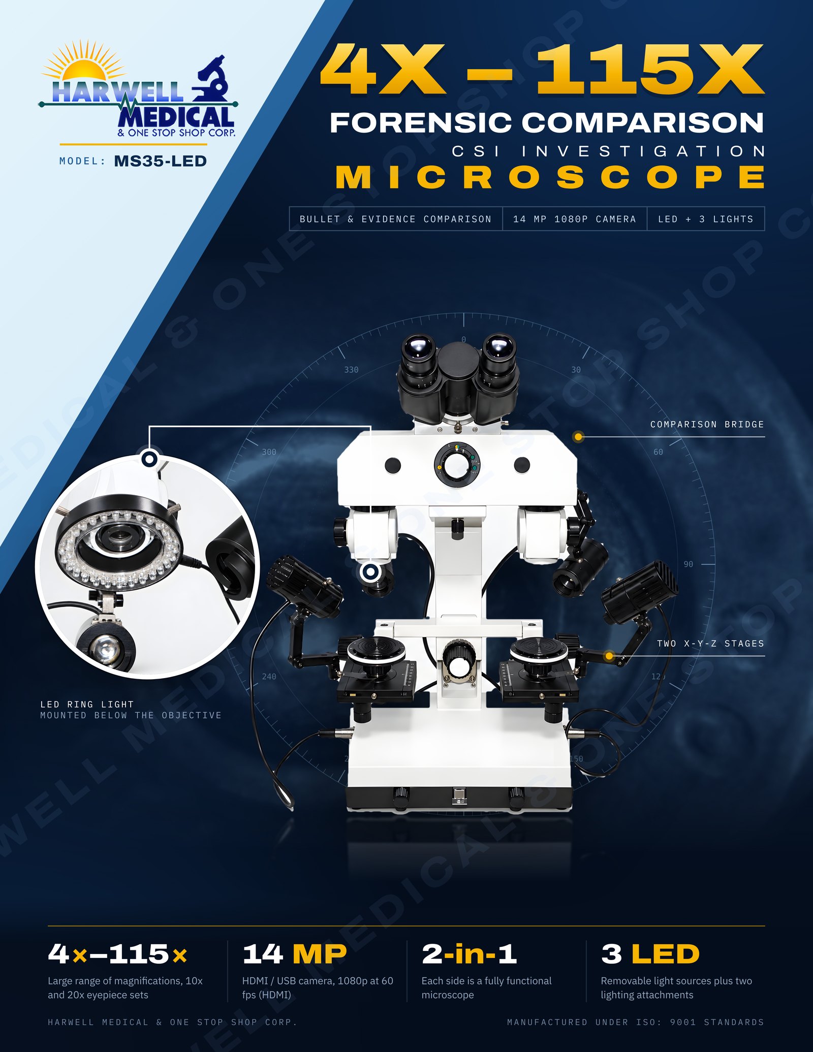
Model: MS35-LED
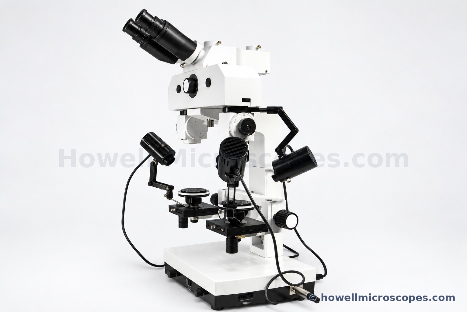
Forensic Comparison Microscope
01Microscope Product Overview
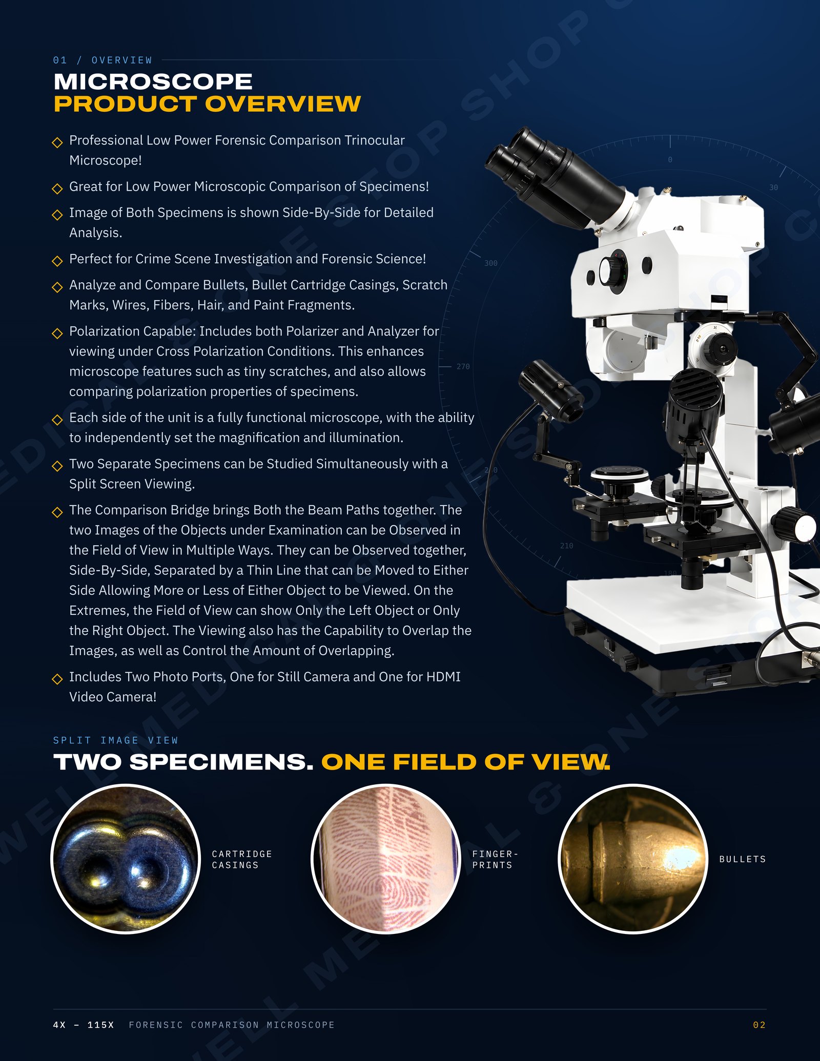
Professional Low Power Forensic Comparison Trinocular Microscope!
Great for Low Power Microscopic Comparison of Specimens!
Image of Both Specimens is shown Side-By-Side for Detailed Analysis.
Perfect for Crime Scene Investigation and Forensic Science!
Analyze and Compare Bullets, Bullet Cartridge Casings, Scratch Marks, Wires, Fibers, Hair, and Paint Fragments.
Polarization Capable: Includes both Polarizer and Analyzer for viewing under Cross Polarization Conditions. This enhances microscope features such as tiny scratches, and also allows comparing polarization properties of specimens.
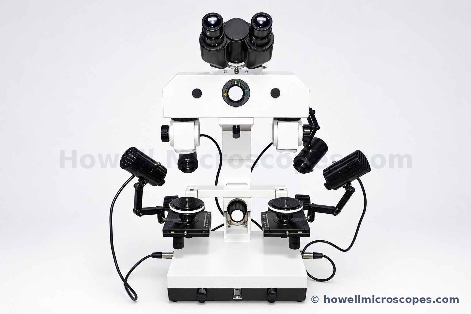
Each side of the unit is a fully functional microscope, with the ability to independently set the magnification and illumination.
Two Separate Specimens can be Studied Simultaneously with a Split Screen Viewing.
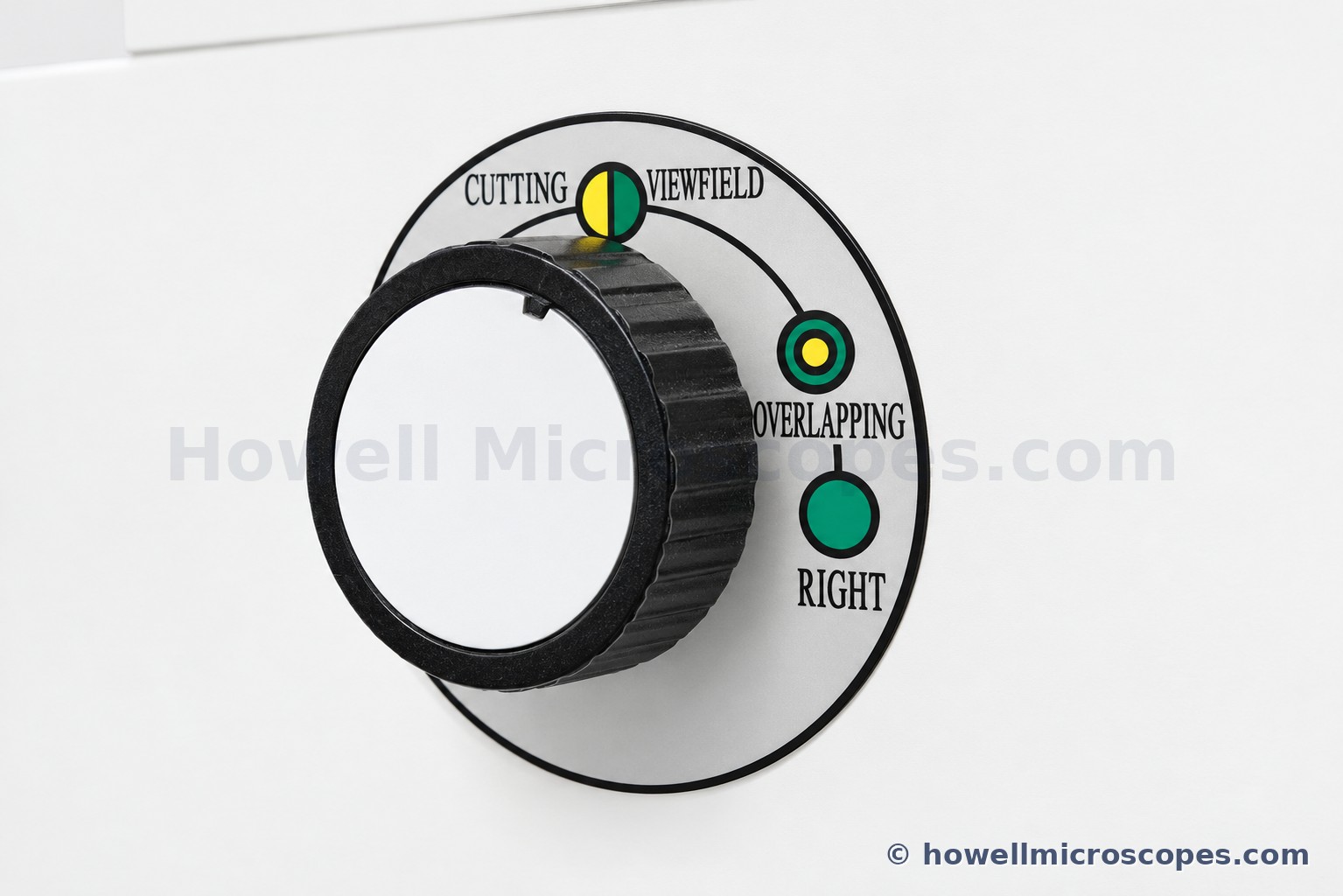
The Comparison Bridge brings Both the Beam Paths together. The two Images of the Objects under Examination can be Observed in the Field of View in Multiple Ways. They can be Observed together, Side-By-Side, Separated by a Thin Line that can be Moved to Either Side Allowing More or Less of Either Object to be Viewed. On the Extremes, the Field of View can show Only the Left Object or Only the Right Object. The Viewing also has the Capability to Overlap the Images, as well as Control the Amount of Overlapping.
Includes Two Photo Ports, One for Still Camera and One for HDMI Video Camera!
02Video and Photography
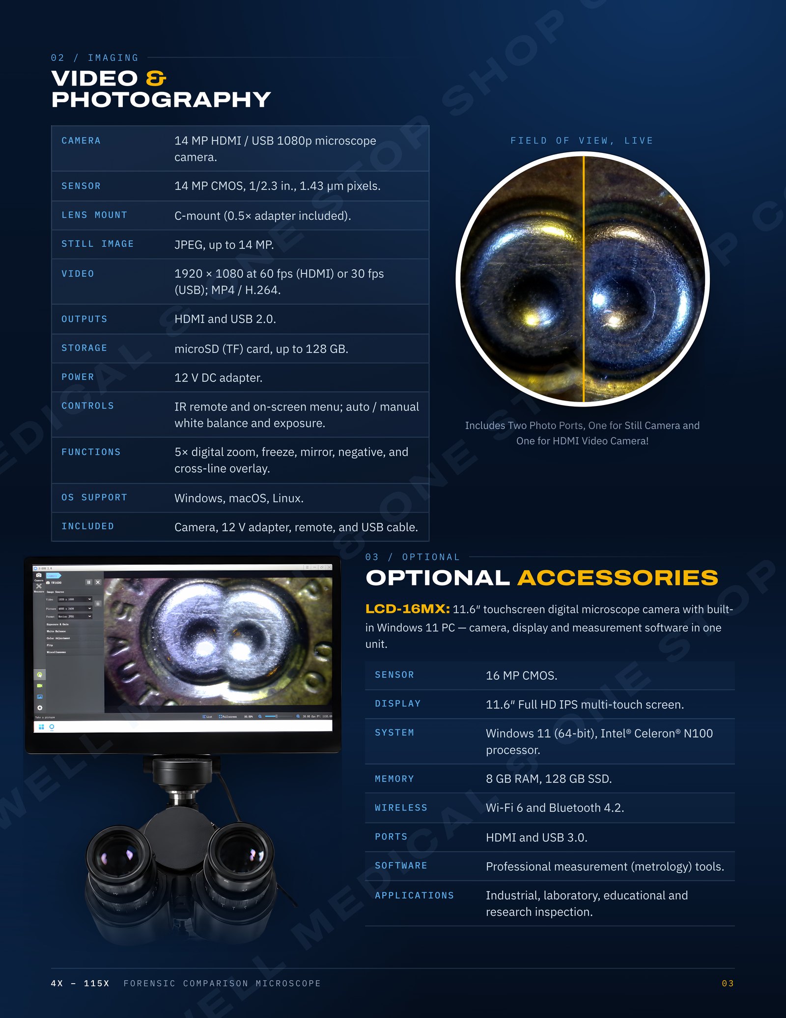
Camera: 14 MP HDMI / USB 1080p microscope camera.
Sensor: 14 MP CMOS, 1/2.3 in., 1.43 µm pixels.
Lens Mount: C-mount (0.5× adapter included).
Still Image: JPEG, up to 14 MP.
Video: 1920 × 1080 at 60 fps (HDMI) or 30 fps (USB); MP4 / H.264.
Outputs: HDMI and USB 2.0.
Storage: microSD (TF) card, up to 128 GB.
Power: 12 V DC adapter.
Controls: IR remote and on-screen menu; auto / manual white balance and exposure.
Functions: 5× digital zoom, freeze, mirror, negative, and cross-line overlay.
OS Support: Windows, macOS, Linux.
Included: Camera, 12 V adapter, remote, and USB cable.
03Optional Accessories/ This LCD Monitor is not Included
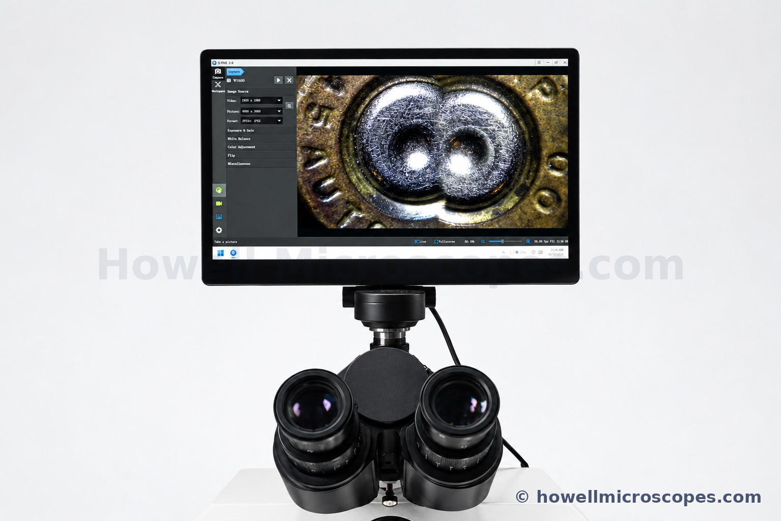
LCD-16MX: 11.6″ touchscreen digital microscope camera with built-in Windows 11 PC — camera, display and measurement software in one unit.
Sensor: 16 MP CMOS.
Display: 11.6″ Full HD IPS multi-touch screen.
System: Windows 11 (64-bit), Intel® Celeron® N100 processor.
Memory: 8 GB RAM, 128 GB SSD.
Wireless: Wi-Fi 6 and Bluetooth 4.2.
Ports: HDMI and USB 3.0.
Software: Professional measurement (metrology) tools.
Applications: Industrial, laboratory, educational and research inspection.
04Eyepieces and Magnification
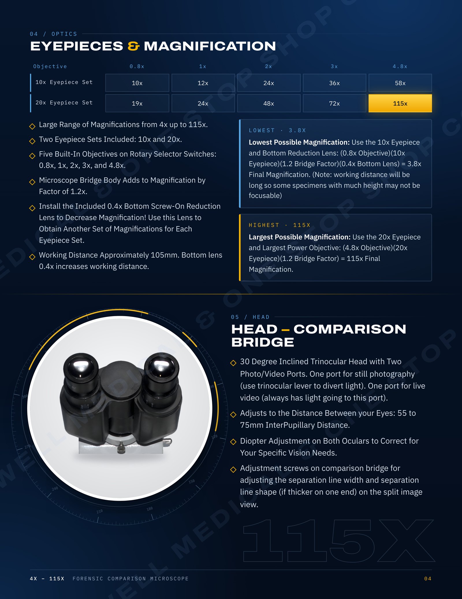
Large Range of Magnifications from 4x up to 115x.
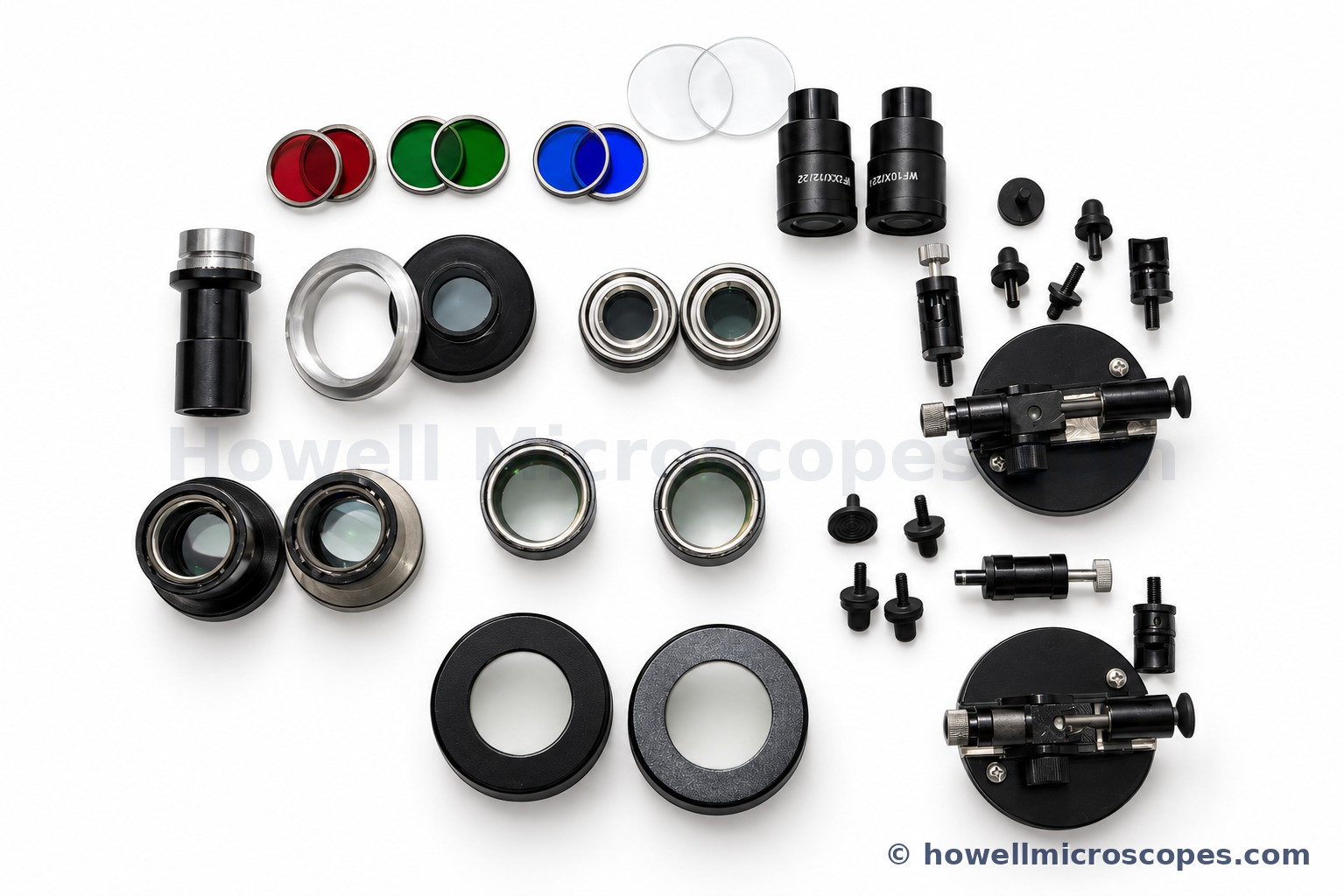
Two Eyepiece Sets Included: 10x and 20x.
Five Built-In Objectives on Rotary Selector Switches: 0.8x, 1x, 2x, 3x, and 4.8x.
Microscope Bridge Body Adds to Magnification by Factor of 1.2x.
10x Eyepiece Set Magnifications: 10x, 12x, 24x, 36x, 58x.
20x Eyepiece Set Magnifications: 19x, 24x, 48x, 72x, 115x.
Install the Included 0.4x Bottom Screw-On Reduction Lens to Decrease Magnification! Use this Lens to Obtain Another Set of Magnifications for Each Eyepiece Set.
Lowest Possible Magnification: Use the 10x Eyepiece and Bottom Reduction Lens: (0.8x Objective)(10x Eyepiece)(1.2 Bridge Factor)(0.4x Bottom Lens) = 3.8x Final Magnification. (Note: working distance will be long so some specimens with much height may not be focusable)
Largest Possible Magnification: Use the 20x Eyepiece and Largest Power Objective: (4.8x Objective)(20x Eyepiece)(1.2 Bridge Factor) = 115x Final Magnification.
Working Distance Approximately 105mm. Bottom lens 0.4x increases working distance.
05Illumination
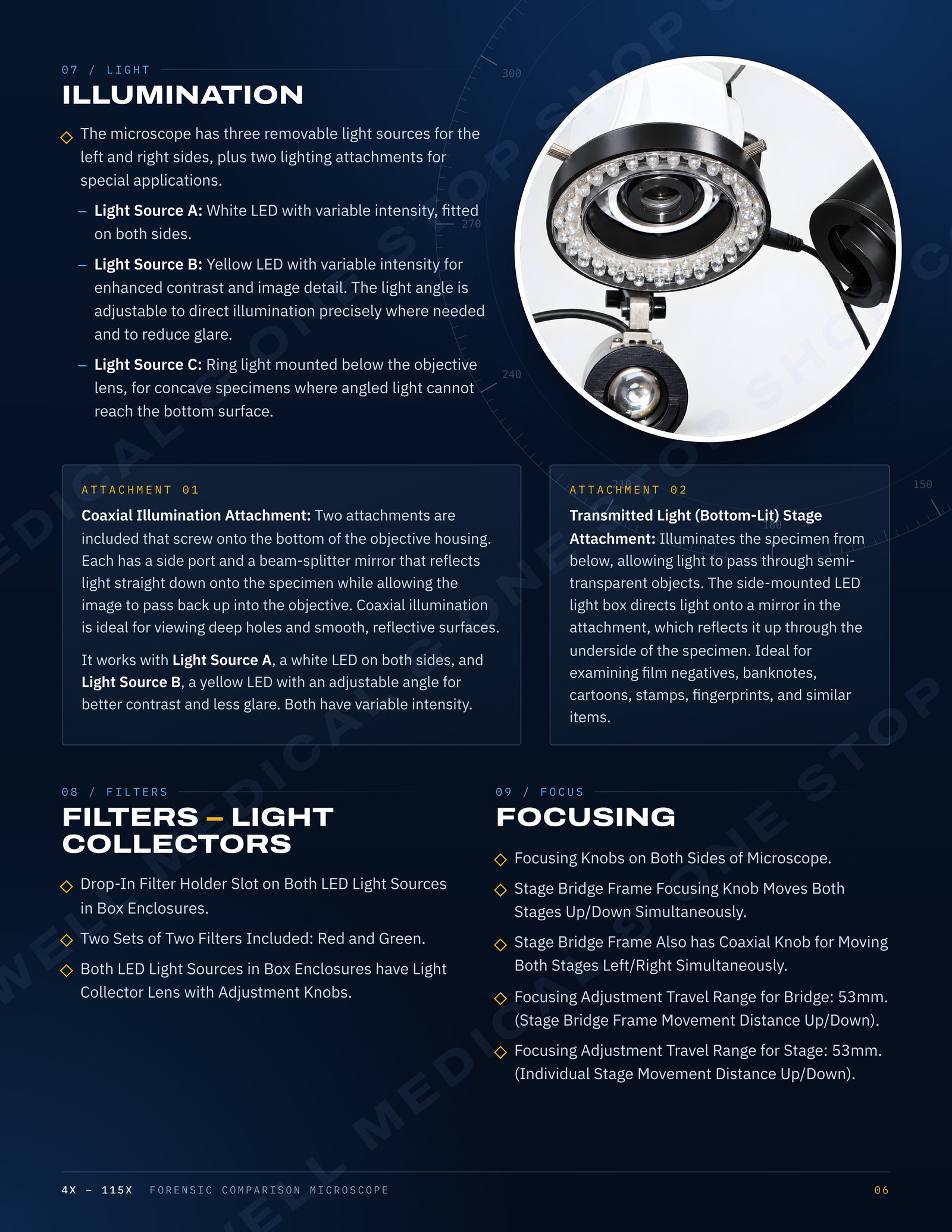
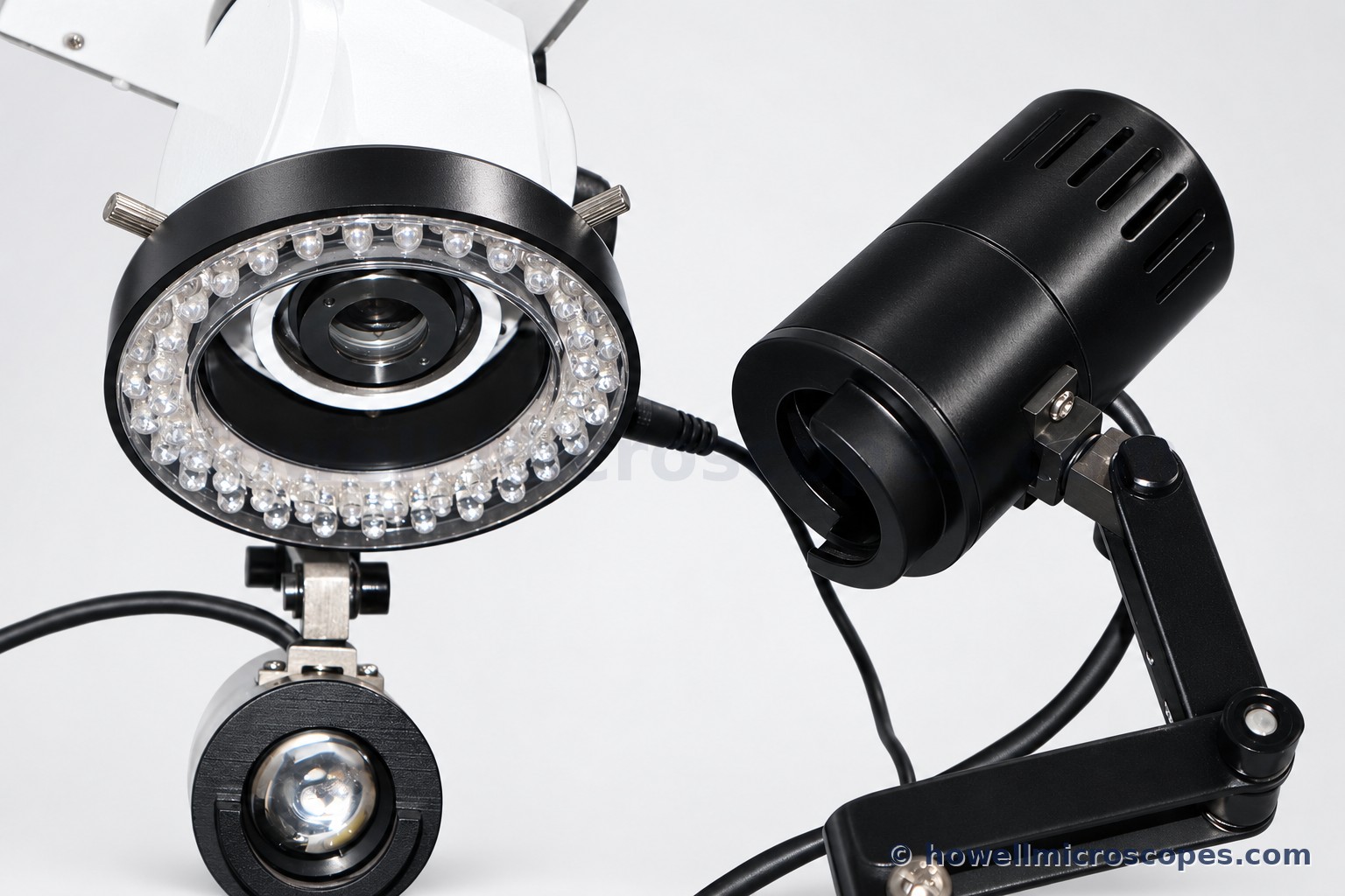
The microscope has three removable light sources for the left and right sides, plus two lighting attachments for special applications.
Light Source A: White LED with variable intensity, fitted on both sides.
Light Source B: Yellow LED with variable intensity for enhanced contrast and image detail. The light angle is adjustable to direct illumination precisely where needed and to reduce glare.
Light Source C: Ring light mounted below the objective lens, for concave specimens where angled light cannot reach the bottom surface.
Coaxial Illumination Attachment: Two attachments are included that screw onto the bottom of the objective housing. Each has a side port and a beam-splitter mirror that reflects light straight down onto the specimen while allowing the image to pass back up into the objective. Coaxial illumination is ideal for viewing deep holes and smooth, reflective surfaces.
It works with Light Source A, a white LED on both sides, and Light Source B, a yellow LED with an adjustable angle for better contrast and less glare. Both have variable intensity.
Transmitted Light (Bottom-Lit) Stage Attachment: Illuminates the specimen from below, allowing light to pass through semi-transparent objects. The side-mounted LED light box directs light onto a mirror in the attachment, which reflects it up through the underside of the specimen. Ideal for examining film negatives, banknotes, cartoons, stamps, fingerprints, and similar items.
06Head – Comparison Bridge
30 Degree Inclined Trinocular Head with Two Photo/Video Ports. One port for still photography (use trinocular lever to divert light). One port for live video (always has light going to this port).
Adjusts to the Distance Between your Eyes: 55 to 75mm InterPupillary Distance.
Diopter Adjustment on Both Oculars to Correct for Your Specific Vision Needs.
Adjustment screws on comparison bridge for adjusting the separation line width and separation line shape (if thicker on one end) on the split image view.
07Comparison Viewing Modes
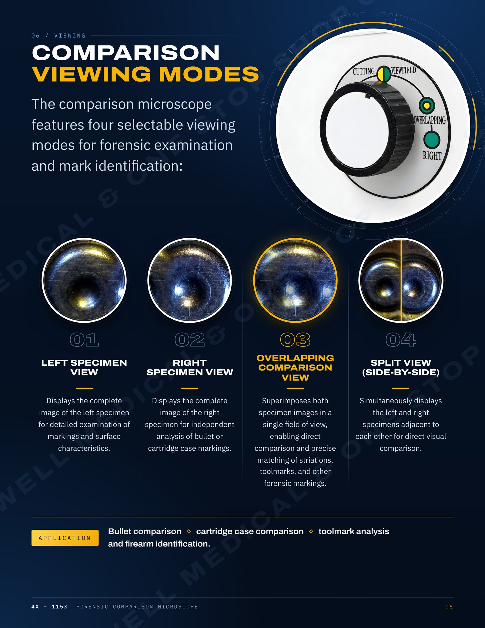
The comparison microscope features four selectable viewing modes for forensic examination and mark identification:
Left Specimen View: Displays the complete image of the left specimen for detailed examination of markings and surface characteristics.
Right Specimen View: Displays the complete image of the right specimen for independent analysis of bullet or cartridge case markings.
Overlapping Comparison View: Superimposes both specimen images in a single field of view, enabling direct comparison and precise matching of striations, toolmarks, and other forensic markings.
Split View (Side-by-Side): Simultaneously displays the left and right specimens adjacent to each other for direct visual comparison.
Application: Bullet comparison, cartridge case comparison, toolmark analysis, and firearm identification.
08Filters – Light Collectors
Drop-In Filter Holder Slot on Both LED Light Sources in Box Enclosures.
Two Sets of Two Filters Included: Red and Green.
Both LED Light Sources in Box Enclosures have Light Collector Lens with Adjustment Knobs.
09Stage Specifications
Two Fully Rotatable Mechanical X-Y-Z Movable Stages, 65mm Diameter with Graduation Marks every 2 Degrees for the Full 360 Degrees.
X-Y Stage Movement Knobs – Range of Movement: 51mm (X-Direction) x 51mm (Y-Direction) x 54mm (Z-Direction).
Rack and Pinion Steel Gears with Knobs for Both X and Y Movements.
Ball Socketed Inclinable Stages: Ball Socket Mounted Stages Provides Capability to Incline.
Vice Type Mechanical Holder Attachments: Great for Holding Objects of Various Diameters, such as Bullets, Coins, and Industrial/Engineering Materials. Object can be Rotated as well as Inclined for Ease of Examining the Surfaces. Sits Directly on Stage. Two Included, One for Each Stage.
10Focusing
Focusing Knobs on Both Sides of Microscope.
Stage Bridge Frame Focusing Knob Moves Both Stages Up/Down Simultaneously.
Stage Bridge Frame Also has Coaxial Knob for Moving Both Stages Left/Right Simultaneously.
Focusing Adjustment Travel Range for Bridge: 53mm. (Stage Bridge Frame Movement Distance Up/Down).
Focusing Adjustment Travel Range for Stage: 53mm. (Individual Stage Movement Distance Up/Down).
11Frame – Base – Size – Weight
Total Overall Height of Microscope: 625mm (top of eyepieces).
12Included Items
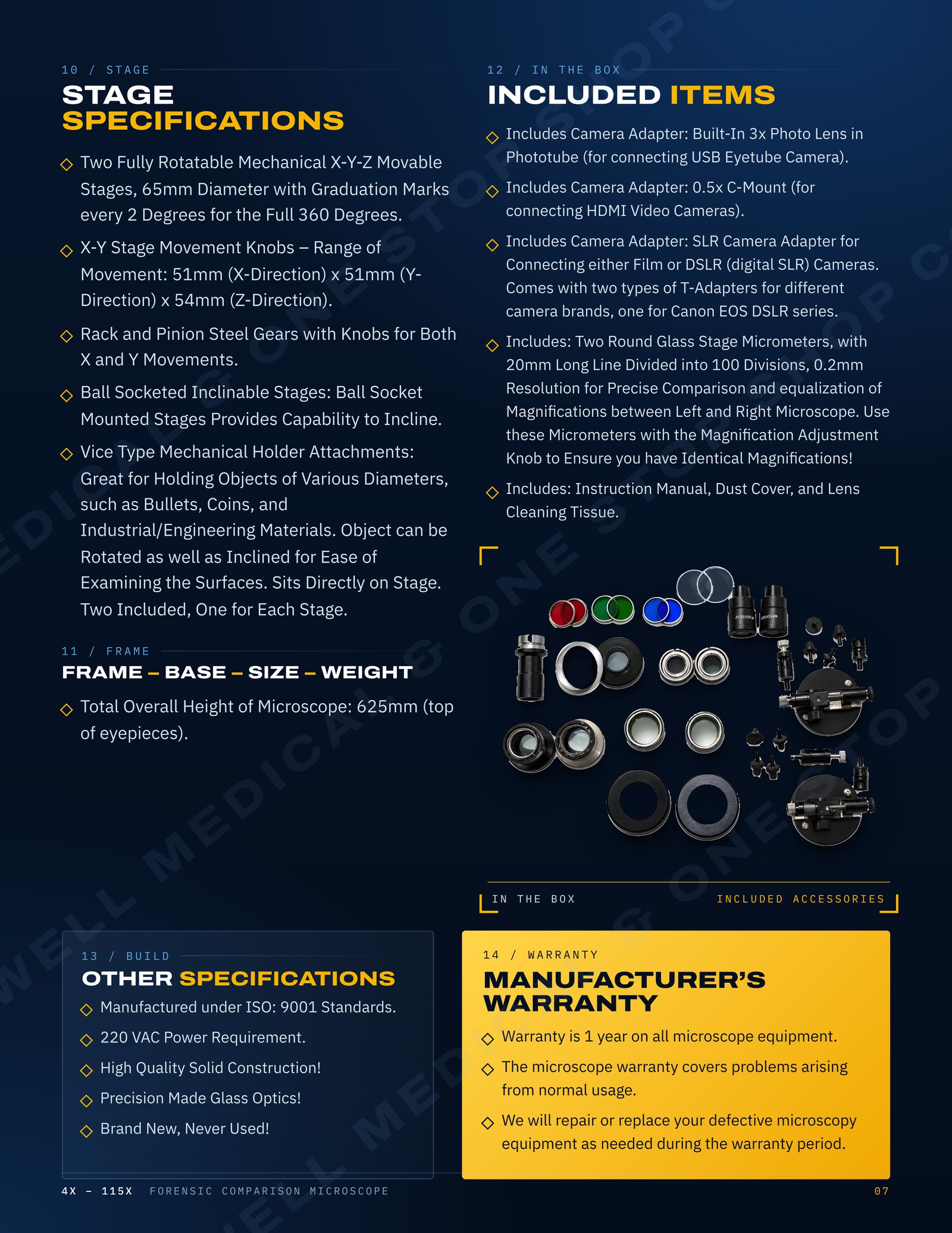
Includes Camera Adapter: Built-In 3x Photo Lens in Phototube (for connecting USB Eyetube Camera).
Includes Camera Adapter: 0.5x C-Mount (for connecting HDMI Video Cameras).
Includes Camera Adapter: SLR Camera Adapter for Connecting either Film or DSLR (digital SLR) Cameras. Comes with two types of T-Adapters for different camera brands, one for Canon EOS DSLR series.
Includes: Two Round Glass Stage Micrometers, with 20mm Long Line Divided into 100 Divisions, 0.2mm Resolution for Precise Comparison and equalization of Magnifications between Left and Right Microscope. Use these Micrometers with the Magnification Adjustment Knob to Ensure you have Identical Magnifications!
Includes: Instruction Manual, Dust Cover, and Lens Cleaning Tissue.
13Other Specifications
Manufactured under ISO: 9001 Standards.
220 VAC Power Requirement.
High Quality Solid Construction!
Precision Made Glass Optics!
Brand New, Never Used!
14Manufacturer’s Warranty
Warranty is 1 year on all microscope equipment.
The microscope warranty covers problems arising from normal usage.
We will repair or replace your defective microscopy equipment as needed during the warranty period.