Product Description
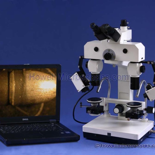
Video and Photography
• High Resolution 3.1 MegaPixel Digital Camera System.
• Complete Digital Microscopy Solution Included.
• Capture high resolution digital microscope images, 2048×1536 pixels.
• View and record full motion live video microscope images. Frame size options for video mode: 2048X1536 (up to 11 FPS, depending on PC), 1024 X 768, 640 X 480, 512 X 384.
• Computer connected digital microscope camera connects via USB2.0. Includes USB cable and MS Windows software.
• Color DSP built into camera provides sharp quality color images. Image sensor size is half inch.
• Includes measuring capability with the software.
Eyepieces and Magnification
• Large Range of Magnifications from 4x up to 115x.
• Two Eyepiece Sets Included: 10x and 20x.
• Five Built-In Objectives on Rotary Selector Switchs: 0.8x, 1x, 2x, 3x, and 4.8x.
• Microscope Bridge Body Adds to Magnification by Factor of 1.2x.
• 10x Eyepiece Set Magnifications: 10x, 12x, 24x, 36x, 58x.
• 20x Eyepiece Set Magnifications: 19x, 24x, 48x, 72x, 115x.
• Install the Included 0.4x Bottom Screw-On Reduction Lens to Decrease Magnification! Use this Lens to Obtain Another Set of Magnifications for Each Eyepiece Set.
• Lowest Possible Magnification: Use the 10x Eyepiece and Bottom Reduction Lens: (0.8x Objective)(10x Eyepiece)(1.2 Bridge Factor)(0.4x Bottom Lens) = 3.8x Final Magnification. (Note: working distance will be long so some specimens with much height may not be focusable)
• Largest Possible Magnification: Use the 20x Eyepiece and Largest Power Objective: (4.8x Objective)(20x Eyepiece)(1.2 Bridge Factor) = 115x Final Magnification.
• Working Distance Approximately 105mm. Bottom lens 0.4x increases working distance.
Illumination
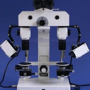
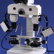
• Two Different Light Source Types for Both the Left and the Right Microscope.
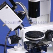
• Light Source A: Variable Intensity 50W Halogen Light Source in Box Enclosure.
• Mounted to Segmented Arm with Capability to Illuminate from Various Positions and Angles.
• This Light Source has Two Separate Locations where it can be Mounted for Maximum Versatility. Mount it Above the Objectives or on the Bracket Extending from the Stage.
• Includes Mini Built-In Fan for Cooling Light Source.
• This Light Source has the Capability to provide Polarized Light with the Built-In Filter Holder.
• Light Source B: Variable Intensity Fiber Optic Light Source. Lamp Type 12V/50W Halogen.
• Bendable Fiber Optic Cables Deliver Illumination Precisely Where Needed. Adjust the Angle as Needed for Glare Reduction.
• Coaxial Illumination Attachment: Includes Two Attachments that Screw to Bottom of Objective Housing. Has Opening on Side to Insert the Tip of the Fiber Optic Cable and a Special Mirror that Reflects the Light Down to Specimen Providing Coaxial Illumination. The Mirror Allows Light from the Specimen to Pass through it and into the Objective. Coaxial Illumination can be Beneficial for Viewing Deep Holes and also Smooth Surfaces.
• Transmitted Light (Bottom Lighted) Stage Attachment: Includes a Special Device that Provides Lighting Under the Specimen. Also Known as Transmitted Light Illumination since can be Transmitted through Semi-Translucent Objects. Great for Examining Film Negatives, Currency Notes, Cartoons, Stamps, Fingerprints, Etc. The side mounted halogen box-light source is directed towards the mirror on the attachment, reflecting the light to the bottom of the specimen to be compared.
Head – Comparison Bridge
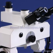
• 30 Degree Inclined Trinocular Head with Two Photo/Video Ports. One port for still photography (use trinocular lever to divert light). One port for live video (always has light going to this port).
• Adjusts to the Distance Between your Eyes: 55 to 75mm InterPupillary Distance.
• Diopter Adjustment on Both Oculars to Correct for Your Specific Vision Needs.
• Adjustment screws on comparison bridge for adjusting the separation line width and separation line shape (if thicker on one end) on the split image view.
Filters – Light Collectors
• Drop-In Filter Holder Slot on Both Halogen Light Sources in Box Enclosures.
• Two Sets of Two Filters Included: Red and Green.
• Both Halogen Light Sources in Box Enclosures have Light Collector Lens with Adjustment Knobs.
Stage Specifications
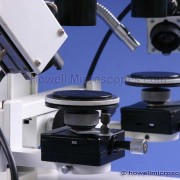
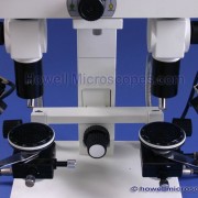
• Two Fully Rotatable Mechanical X-Y-Z Movable Stages, 65mm Diameter with Graduation Marks every 2 Degrees for the Full 360 Degrees.
• X-Y Stage Movement Knobs – Range of Movement: 51mm (X-Direction) x 51mm (Y-Direction) x 54mm (Z-Direction).
• Rack and Pinion Steel Gears with Knobs for Both X and Y Movements.
• Ball Socketed Inclinable Stages: Ball Socket Mounted Stages Provides Capability to Incline.
• Vice Type Mechanical Holder Attachments: Great for Holding Objects of Various Diameters, such as Bullets, Coins, and Industrial/Engineering Materials. Object can be Rotated as well as Inclined for Ease of Examining the Surfaces. Sits Directly on Stage. Two Included, One for Each Stage.
Focusing
• Focusing Knobs on Both Sides of Microscope.
• Stage Bridge Frame Focusing Knob Moves Both Stages Up/Down Simultaneously.
• Stage Bridge Frame Also has Coaxial Knob for Moving Both Stages Left/Right Simultaneously.
• Focusing Adjustment Travel Range for Bridge: 53mm. (Stage Bridge Frame Movement Distance Up/Down).
• Focusing Adjustment Travel Range for Stage: 53mm. (Individual Stage Movement Distance Up/Down).
Frame – Base – Size – Weight
• Total Overall Height of Microscope: 625mm (top of eyepieces).
Included Items
• Includes Camera Adapter: Built-In 3x Photo Lens in Phototube (for connecting USB Eyetube Camera).
• Includes Camera Adapter: 0.5x C-Mount (for connecting CCD Video Cameras).
• Includes Camera Adapter: SLR Camera Adapter for Connecting either Film or DSLR (digital SLR) Cameras. Comes with two types of T-Adapters for different camera brands, one for Canon EOS DSLR series.
• Includes: Two Round Glass Stage Micrometers, with 20mm Long Line Divided into 100 Divisions, 0.2mm Resolution for Precise Comparison and equalization of Magnifications between Left and Right Microscope. Use these Micrometers with the Magnification Adjustment Knob to Ensure you have Identical Magnifications!
• Includes: Instruction Manual, Dust Cover, Lens Cleaning Tissue, and Two Extra Bulbs.
Other Specifications
• Manufactured under ISO: 9001 Standards.
• 220 VAC Power Requirement.
• High Quality Solid Construction!
• Precision Made Glass Optics!
• Brand New, Never Used!
Manufacturer’s Warranty
• Warranty is 1 years on all microscope equipment.
• The microscope warranty covers problems arising from normal usage.
• We will repair or replace your defective microscopy equipment as needed during the warranty period.

Reviews
There are no reviews yet.